Imaging form unearths unused cells and constructions in human mind tissue
A unused microscopy methodology that permits high-resolution imaging may just one future support medical doctors diagnose and deal with mind tumors.
The use of a brochure microscopy methodology, MIT and Brigham and Girls’s Health center/Harvard Clinical College researchers have imaged human mind tissue in higher component than ever ahead of, revealing cells and constructions that weren’t prior to now visual.
Amongst their findings, the researchers found out that some “low-grade” mind tumors include extra putative competitive tumor cells than anticipated, suggesting that a few of these tumors could also be extra competitive than prior to now concept.
The researchers hope that this system may just in the end be deployed to diagnose tumors, generate extra correct prognoses, and support medical doctors make a selection therapies.
“We’re starting to see how important the interactions of neurons and synapses with the surrounding brain are to the growth and progression of tumors. A lot of those things we really couldn’t see with conventional tools, but now we have a tool to look at those tissues at the nanoscale and try to understand these interactions,” says Pablo Valdes, a former MIT postdoc who’s now an laborer schoolmaster of neuroscience on the College of Texas Clinical Segment and the supremacy writer of the find out about.
Edward Boyden, the Y. Eva Tan Educator in Neurotechnology at MIT; a schoolmaster of organic engineering, media arts and sciences, and mind and cognitive sciences; a Howard Hughes Clinical Institute investigator; and a member of MIT’s McGovern Institute for Mind Analysis and Koch Institute for Integrative Most cancers Analysis; and E. Antonio Chiocca, a schoolmaster of neurosurgery at Harvard Clinical College and chair of neurosurgery at Brigham and Girls’s Health center, are the senior authors of the find out about, which seems lately in Science Translational Medication.
Making molecules visual
The unused imaging form is in response to growth microscopy , a method evolved in Boyden’s lab in 2015 in response to a easy premise: In lieu of the usage of robust, dear microscopes to acquire high-resolution photographs, the researchers devised a solution to amplify the tissue itself, permitting it to be imaged at very tall decision with a common shiny microscope.
The methodology works via embedding the tissue right into a polymer that swells when aqua is added, and upcoming softening up and breaking aside the proteins that in most cases keep tissue in combination. After, including aqua swells the polymer, pulling all of the proteins except each and every alternative. This tissue expansion lets in researchers to acquire photographs with a decision of round 70 nanometers, which was once prior to now imaginable most effective with very specialised and costly microscopes comparable to scanning electron microscopes.
In 2017, the Boyden lab evolved a solution to amplify guarded human tissue specimens, however the chemical reagents that they old additionally destroyed the proteins that the researchers have been curious about labeling. By way of labeling the proteins with fluorescent antibodies ahead of growth, the proteins’ location and identification might be visualized then the growth procedure was once whole. Then again, the antibodies most often old for this sort of labeling can’t simply squeeze via densely packed tissue ahead of it’s expanded.
So, for this find out about, the authors devised a unique tissue-softening protocol that breaks up the tissue however preserves proteins within the pattern. Then the tissue is expanded, proteins will also be labelled with commercially to be had fluorescent antibodies. The researchers upcoming can carry out a number of rounds of imaging, with 3 or 4 other proteins categorised in each and every spherical. This labelling of proteins allows many extra constructions to be imaged, as a result of as soon as the tissue is expanded, antibodies can squeeze via and label proteins they couldn’t prior to now succeed in.
“We open up the space between the proteins so that we can get antibodies into crowded spaces that we couldn’t otherwise,” Valdes says. “We saw that we could expand the tissue, we could decrowd the proteins, and we could image many, many proteins in the same tissue by doing multiple rounds of staining.”
Running with MIT Worker Educator Deblina Sarkar, the researchers demonstrated a mode of this “decrowding” in 2022 the usage of mouse tissue.
The unused find out about ended in a decrowding methodology for importance with human mind tissue samples which might be old in scientific settings for pathological prognosis and to lead remedy choices. Those samples will also be tougher to paintings with as a result of they’re generally embedded in paraffin and handled with alternative chemical substances that wish to be damaged unwell ahead of the tissue will also be expanded.
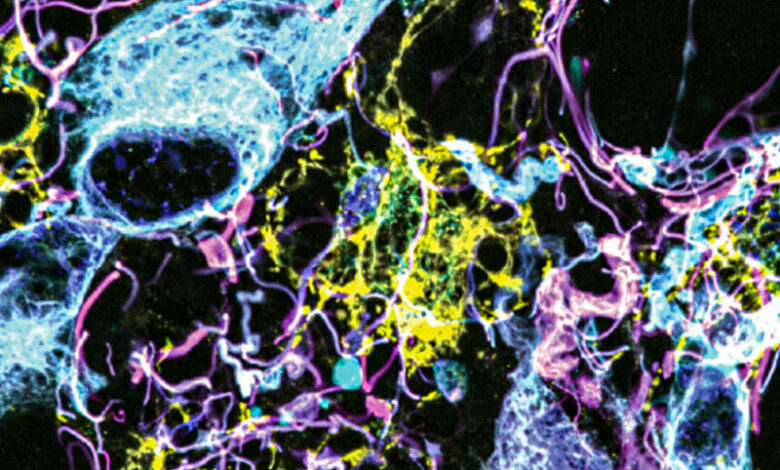
On this find out about, the researchers categorised as much as 16 other molecules in step with tissue pattern. The molecules they centered come with markers for numerous constructions, together with axons and synapses, in addition to markers that determine cellular varieties comparable to astrocytes and cells that mode blood vessels. Additionally they categorised molecules related to tumor aggressiveness and neurodegeneration.
The use of this way, the researchers analyzed wholesome mind tissue, at the side of samples from sufferers with two forms of glioma – high-grade glioblastoma, which is probably the most competitive number one mind tumor, with a unpriviledged diagnosis, and low-grade gliomas, which might be regarded as much less competitive.
“We wanted to look at brain tumors so that we can understand them better at the nanoscale level, and by doing that, to be able to develop better treatments and diagnoses in the future. At this point, it was more developing a tool to be able to understand them better, because currently in neuro-oncology, people haven’t done much in terms of super-resolution imaging,” Valdes says.
A diagnostic instrument
To spot competitive tumor cells in gliomas they studied, the researchers categorised vimentin, a protein this is present in extremely competitive glioblastomas. To their miracle, they discovered many extra vimentin-expressing tumor cells in low-grade gliomas than have been distinguishable the usage of any alternative form.
“This tells us something about the biology of these tumors, specifically, how some of them probably have a more aggressive nature than you would suspect by doing standard staining techniques,” Valdes says.
When glioma sufferers go through surgical procedure, tumor samples are guarded and analyzed the usage of immunohistochemistry staining, which is able to disclose positive markers of aggressiveness, together with probably the most markers analyzed on this find out about.
“These are incurable brain cancers, and this type of discovery will allow us to figure out which cancer molecules to target so we can design better treatments. It also proves the profound impact of having clinicians like us at the Brigham and Women’s interacting with basic scientists such as Ed Boyden at MIT to discover new technologies that can improve patient lives,” Chiocca says.
The researchers hope their growth microscopy methodology may just permit medical doctors to be informed a lot more about sufferers’ tumors, serving to them to decide how competitive the tumor is and guiding remedy alternatives. Valdes now plans to do a bigger find out about of tumor varieties to struggle to ascertain diagnostic tips in response to the tumor characteristics that may be unmistakable the usage of this system.
“Our hope is that this is going to be a diagnostic tool to pick up marker cells, interactions, and so on, that we couldn’t before,” he says. “It’s a practical tool that will help the clinical world of neuro-oncology and neuropathology look at neurological diseases at the nanoscale like never before, because fundamentally it’s a very simple tool to use.”
Boyden’s lab additionally plans to importance this option to find out about alternative sides of mind serve as, in wholesome and diseased tissue.
“Being able to do nanoimaging is important because biology is about nanoscale things – genes, gene products, biomolecules – and they interact over nanoscale distances,” Boyden says. “We can study all sorts of nanoscale interactions, including synaptic changes, immune interactions, and changes that occur during cancer and aging.”
Paper: “Improved immunostaining of nanostructures and cells in human brain specimens through expansion-mediated protein decrowding”


